
In-Situ And 4d Characterization
3D microstructural information from samples that incrementally undergo changes
- Non-destructive characterization of the 3D microstructure of materials under controlled perturbations
- Observe the evolution of structures over time
- Resolution at a Distance ensures the highest resolution across large working distances, accommodating both sample, environmental chamber, and high precision in-situ load rigs without sacrificing resolution
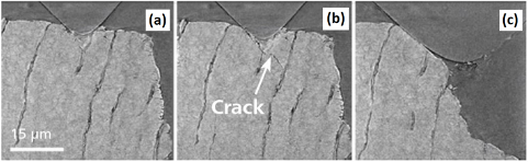
Sample of elephant dentin mounted in the nanomechanical test stage. Evolution of crack propagation. (a) starting configuration, in (b) a crack is forming and in (c) a piece of dentine has fractured off. (Source: B. Hornberger, H. Bale and A. Merkle, "X-ray microscopy for in situ characterization of 3D nanostructural evolution in the laboratory," X-Ray Nanoimaging: Instruments and Methods II, vol. 9592, 2015).
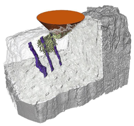
3D rendering showing the morphology of a selected crack (green) in relation to neighboring tubules (blue) and the indenter tip (orange) of the elephant dentine sample. (Source: B. Hornberger, H. Bale and A. Merkle, "X-ray microscopy for in situ characterization of 3D nanostructural evolution in the laboratory," X-Ray Nanoimaging: Instruments and Methods II, vol. 9592, 2015).